Saturday, October 15, 2022

                                                                            Virology


Genome


The Cache Valley virus genome is split into three parts. The three parts are called the small, medium, and large segments, based on the number of bases. The large segment encodes the L protein, which is the RNA dependent RNA polymerase. The small segment utilizes an open reading frame with alternative initiation sites to encode two proteins. Depending on the initiation site, it can either code for the protein that makes up the nucleocapsid, N, or a non-structural protein, NSs. The medium segment encodes 2 type 1 integral transmembrane glycoproteins, Gn and Gc, as well as a non-structural protein Nsm. The Gc and Gn proteins start as one precursor protein and are then cleaved cotranslationally. They are modified by N-linked glycosylation.


Replication cycle

The attachment, entry, replication, and release of CVV specifically have not been studied. However, there is information of the replication cycle for the genus orthobunyavirus, which CVV is a part of. A heterodimer of integral transmembrane proteins Gn and Gc form spikes on the surface of the virus particle. They are involved in virus attachment and cell fusion.  Once inside the cell, the viral membrane fuses with the endosomal membrane, and the virus genome is released. Transcription involves an RNA dependent RNA polymerase, and it occurs in the cytoplasm of the cell. Transcription of the tripartite genome is terminated by a strong hairpin loop sequence at the end of each segment. Once the virus has replicated enough, it is encapsidated.   Assembly and budding of the newly synthesized virions occurs at the membranes of the Golgi apparatus. 

Host interactions

In terms of CVV alone, very little is known about the regulation of host-processes and interactions with host cells. However, CVVs Bunyamwera serogroup's two non-structural proteins play an important role in infection. Bunyamwera virus (BUNV) codes for two non-structural proteins: NSm on the medium RNA segment and NSs on the smallest RNA segment.[10] Bunyamwera virus NSs protein is a nonessential gene that contributes to viral pathogenesis. It has been shown that in mammalian cells, NSs induces shut-off of host protein synthesis, which leads to cell death. It also counteracts the host cell antiviral response and seems to be the main virulence factor, acting at the level of transcription by inhibiting RNA polymerase II–mediated transcription. In mosquito cells neither host cell transcription nor translation are inhibited, and although so far no function for the orthobunyavirus NSs protein has been found in mosquito cells, it seems the differential behavior of NSs could be one of the factors responsible for different outcomes of infection in mammalian and mosquito cell lines.


Human cases

Prior to 1956 there were no known cases of acute infections of Cache Valley virus (CVV) in humans. However antibodies against CVV have been reported.[ One study found neutralizing antibody to CVV in 12% of 356 persons surveyed in Maryland and Virginia in the 1960s. These results and other such serosurveys are based on nonrandom sampling and therefore often difficult to interpret.


CVV disease is a neuroinvasive illness.  Of the three confirmed human cases of CVV disease two resulted in non-fatal meningitis, only the first case caused fatal encephalitis and multiorgan faukyre,


The first case was a 28-year-old man from North Carolina in 1995. It is likely he was infected with the virus via mosquitos during a deer-hunting trip. The patient's first symptoms were muscle pain, fever, chills and a headache. He began vomiting the day after the first symptoms appeared. Six days after the onset of the illness more severe symptoms appeared including confusion, tachycardia (elevated heart rate), a rash, bilateral conjunctivitis and meningismus. The next day the patient became hypotensive and delirious. Later respiratory failure, seizures and necrosis of the fingers and toes occurred. One leg was amputated because of extensive muscle and cutaneous necrosis. Seven months after the onset of the illness the patient died of pulmonary complications. The causative agent was identified as a virus in the family Bunyaviridae by electron microscopy. This was then identified genetically as CVV. Viremia was recorded seven days after the onset of fever; this is a longer period of viremia than what is normally observed in cases of Bunyaviridae infections.


The second human case of CVV was a 41-year-old man from Wisconsin in October 2003. He developed an acute illness with severe nausea, vomiting, fatigue and headache. He was diagnosed with acute aseptic meningitis. After three days the patient was released from the hospital; he reported feeling fully recovered four months later, though he experienced headaches more frequently than usual. The causative agent was observed with electron microscopy as being virions morphologically similar to bunyaviruses. Nucleotide sequencing identified the virus as CVV.


The third human case of CVV was a 63-year-old woman in New York, in September 2011. When she was admitted to the hospital her symptoms were fever, headache, neck stiffness and photophobia. A week before she had noticed a lesion on her arm, as this began to fade a rash developed and spread. She then developed a fever and symptoms of meningitis. The patient was discharged, but returned the next day with nausea and vomiting. She was diagnosed with aseptic meningitis. She was discharged four days later. Two months after this she reported ongoing difficulties in word finding and headaches. CVV was identified as the causative agent by PCR, sequence analysis confirmed this identification.


It is likely that CVV disease is underreported. Very few human cases have been reported despite its wide geographic distribution and the large number of mosquito species that transmit it.[4] The rarity of CVV disease diagnosis is partly due to the fact that laboratories rarely test for CVV. Therefore, the true incidence of CVV disease and its full clinical range are still unknown.  Given the widespread distribution of CVV and other viruses in the same serogroup in the United States it is possible some unexplained cases of severe multiorgan failure, congenital anomalies and human viral encephalitis may be due to CVV or similar viruses. More research of such cases is needed.


Other animals

Cache Valley virus is the most common Orthobunyavirus in North America,[17] and while isolated in 1956, was only linked to disease in Texas in 1987 during a large occurrence of aborted and malformed lambs in a sheep flock. The virus does not only infect sheep, however, as In 2002 a survey conducted in 22 states showed 28% of cattle expressed specific antibodies to CVV.  Cache Valley virus has also been identified as a cause of fetal abnormalities in goats. Other serological surveys have also shown antibodies to CVV in domestic and wild ruminants, along with horses. Of wild ruminants, deer have a very high seroprevalence. With viraemia lasting 1 to 3 days, they are easily able to spread the virus to vectors including Culicoides midges and Aedes, Anopheles, Coquillettidia and Culiseta group mosquitoes. Therefore, deer tend to act as amplifying hosts to the virus.


Symptoms in sheep

While the virus is able to replicate in adult animals, besides a slight febrile response in some cases, there are no known symptoms of infection.  There is a quick period of viraemia before seroconversion and the infection is cleared quickly by the animal's immune system. However, if the animal is pregnant and not protected by antibodies from a previous infection, Cache Valley virus can be very lethal to a developing fetus.


The symptoms the fetus develops from CVV infection are largely age dependent. At less than 28 days of gestation, the embryo usually dies and is reabsorbed by the mother.  Between 28 and 45 days of gestation, infection leads to malformations in the developing fetus and occasionally leads to abortions. Early in this window, between 28 and 36 days, the virus leads to both central nervous system and musculoskeletal defects, while after 36 days of gestation infection only leads to musculoskeletal deformities.   Death of the fetus usually occurs between 27 and 35 days gestation, when the central nervous system tissues are most susceptible. After 45–50 days of gestation CVV infection is not expected to cause harmful effects. After 7 6 days the fetus has a functioning immune system and antibodies to the virus are produced.


Autopsies of infected fetuses show severe lesions in the brain and spinal column, ranging from microscopic in size to whole sections of the brain missing.  In one laboratory case, the cerebral hemispheres were nothing more than fluid-filled sacs that were easily ruptured. The most common musculoskeletal deformations include arthrogryposis and greatly reduced muscle mass, with the most severe cases having torticollis, scoliosis, and kyphosis. While most affected lambs are stillborn, those that do survive are usually so weak they die within minutes of birth.  During the time that they are alive, these amb are reported to act abnormal, such as acting weak, drowsy, or walking unsteadily.


Jan Ricks Jennings, MHA, LFACHE

Senior Consultant

Senior Management Resources, LLC


Jan.Jennings@EagleTalons.Net

JanJenningsBlog.Blogspot.com


412.913.0636 Cell

724.733.0509 Office


October 15, 2022


Sunday, October 9, 2022

                                                              Candida auris


Candida auris can cause invasive candidiasis (fungemia) in which the bloodstream, the central nervous system, and internal organs are infected. It has attracted widespread attention because of its multiple drug resistance. Treatment is also complicated because it is easily misidentified as other Candida species.


Candida auris was first described in 2009 after it was isolated from the ear canal of a 70-year-old Japanese woman at the Tokyo Metropolitan Geriatric Hospital in Japan. In 2011, South Korea saw its first cases of disease-causing C. auris. Reportedly, this spread across Asia and Europe, and first appeared in the U.S. in 2013.


DNA analysis of four distinct but drug-resistant strains of Candida auris indicate an evolutionary divergence taking place at least 4,000 years ago, with a common leap among the four varieties into drug-resistance possibly linked to widespread azole-type antifungal use in agriculture.[4] However, explanations for its emergence remain speculative.


Contents

Identification and morphology

First identified in 2009, Candida auris is a species of ascomycetous fungus of the genus Candida that grows as a yeast. Its name comes from the Latin word for ear, auris.  It forms smooth, shiny, whitish-gray, viscous colonies on growth media. Microscopically cells are ellipsoid in shape.


Clinical significance

Candida auris is one of the few Candida species that can cause candidiasis in humans. Candidiasis is most often acquired in hospitals by patients with weakened immune systems. It can cause invasive candidiasis, in which the blood stream (fungemia), the central nervous system, kidneys, liver, bones, muscles, joints, spleen, or eyes are invaded. It usually presents with other co-morbidities such as diabetes, sepsis, lung diseases, and kidney diseases.


Candida auris has attracted increased clinical attention because of its multiple drug resistance.


In vitro, more than 90% of C. auris isolates are resistant to fluconazole and a range of 3–73% of C. auris isolates are resistant to voriconazole, while other triazoles (posaconazole, itraconazole, and isavuconazole) display better activity.  Of isolates 13% to 35% were reported resistant to amphotericin B;[8][10] however, most isolates are susceptible to echinocandins.


Treatment is complicated because C. auris is easily misidentified as various other Candida species.  A brief outline of its clinical relevance as of 2016, understandable by general audiences, was published by the Center for Infectious Disease Research and Policy at the University of Minnesota.


According to the Centers for Disease Control and Prevention (CDC), 30–60% of people with C. auris bloodstream infections (BSI) have died.   However, many of these people had other serious illnesses and conditions (comorbidities) that also increased their risk of death.


Vaccine development

There is currently no vaccine for Candida auris, however, attempts have been made, with experiments involving the NDV-3A vaccine that successfully immunized mice against the fungus  The vaccine also improved the protective efficacy of the antifungal drug micafungin against C. auris infection in the bloodstream.


Genome

Several draft genomes from whole genome sequencing have been published.[6][18] C. auris has a genome size of 12.3–12.5 Mb with a GC-content of 44.5–44.8%. The C. auris genome was found to encode several genes for the ABC transporter family, a major facilitator superfamily, which helps to explain its multiple drug resistance.[6] Its genome also encodes virulence-related gene families such as lipases, oligopeptide transporters, mannosyl transferases and transcription factors which facilitate colonization, invasion, and iron acquisition. Another factor contributing to antifungal resistance is the presence of a set of genes known to be involved in biofilm formation.[6]


More studies are needed to determine whether the phylogenetic divergence of C. auris clones exhibits region-specific patterns of invasiveness, virulence, and/or drug resistance.


Epidemiology

Geographical differentiation

The phylogenetics of C. auris suggest distinct genotypes exist in different geographical regions with substantial genomic diversity. A variety of sequence-based analytical methods have been used to support this finding.[citation needed.


Whole genome sequencing and analyses of isolates from Pakistan, India, South Africa, Venezuela, Japan, and previously sequenced C. auris genomes deposited in the National Center for Biotechnology Information's Sequence Read Archive. identified a distinct geographic distribution of genotypes.[8] Four distinct clades separated by tens of thousands of single-nucleotide polymorphisms were identified. The distribution of these clades segregated geographically to South Asia (India and Pakistan), South Africa, Venezuela, and Japan with minimal observed intraregion genetic diversity.


Amplified fragment length polymorphism analysis of C. auris isolates from the United Kingdom, India, Japan, South Africa, South Korea, and Venezuela suggested that the London isolates formed a distinct cluster compared to the others.[21]


Comparison of ribosomal DNA sequences of C. auris isolates from Israel, Asia, South Africa, and Kuwait found that the strains from Israel were phylogenetically distinct from those from the other regions.[22] Chatterjee et al. wrote in 2015, "Its actual global distribution remains obscure as the current commercial methods of clinical diagnosis misi


dentify it as C. haemulonii."


History


Candida auris infections, world distribution as of 2019


Map of Candida auris infections in United States as of 2019

Candida auris was first described after it was isolated from the ear canal of a 70-year-old Japanese woman at the Tokyo Metropolitan Geriatric Hospital in Japan. It was isolated based on its ability to grow in the presence of the fungicide micafungin, an echinocandin class fungicide.[1] Phenotypic, chemotaxonomic and phylogenetic analyses established C. auris as a new strain of the genus Candida.


The first three cases of disease-causing C. auris were reported from South Korea in 2011.  Two isolates had been obtained during a 2009 study and a third was discovered in a stored sample from 1996.  All three cases had persistent fungemia, i.e. bloodstream infection, and two of the patients subsequently died due to complications. Notably, the isolates initially were misidentified as Candida haemulonii and Rhodotorula glutinis using standard methods, until sequence analysis correctly identified them as C. auris. These first cases emphasize the importance of accurate species identification and timely application of the correct antifungal for the effective treatment of candidiasis with C. auris.


During 2009–2011, 12 C. auris isolates were obtained from patients at two hospitals in Delhi, India. The same genotype was found in distinct settings: intensive care, surgical, medical, oncologic, neonatal, and pediatric wards, which were mutually exclusive with respect to health care personnel.  Most had persistent candidemia and a high mortality rate was observed. All isolates were of the same clonal strain, however, and were only identified positively by DNA sequence analysis. As previously, the strain was misidentified with established diagnostic laboratory tests. The Indian researchers wrote in 2013 that C. auris was much more prevalent than published reports indicate since most diagnostic laboratories do not use sequence-based methods for strain identification.


The fungus spread to other continents and eventually, a multi-drug-resistant strain was discovered in Southeast Asian countries in early 2016

The first report of a C. auris outbreak in Europe was an October 2016 in Royal Brompton Hospital, a London cardio-thoracic hospital.[21] In April 2017, CDC director Anne Schuchat named it a "catastrophic threat". As of May 2017 the CDC had reported 77 cases in the United States. Of these, 69 were from samples collected in New York and New Jersey.


As of 31 August 2019 the number of cases of people having contracted C. auris in the United States had risen to 806, with 388 reported in New York, 137 in New Jersey and 227 in Illinois, according to the CDC.


Since it was first observed in the United Kingdom, it has spread to more than 20 NHS Trust hospitals and infected 200 people.


As of April 2019, the CDC has documented cases of C. auris from the following countries: Australia, Austria, Belgium, Canada, China, Colombia, France, Germany, India, Iran, Israel, Japan, Kenya, Kuwait, Malaysia, Mexico, the Netherlands, Norway, Oman, Pakistan, Panama, Russia, Saudi Arabia, Singapore, South Africa, South Korea, Spain, Switzerland, Taiwan, Thailand, the United Arab Emirates, the United Kingdom, the United States, and Venezuela.


Candida auris fungus (C. auris) is a multi-drug–resistant fungal infection that spreads in hospitals and is extremely deadly—killing as many as one in three who get it.


—Abby Haglage of Yahoo! Lifestyle,[31] citing the Centers for Disease Control and Prevention.

Arturo Casadevall, MD, PhD, and Molecular Microbiology and Immunology chair at Johns Hopkins Bloomberg School of Public Health stated:


What this study suggests is this is the beginning of fungi adapting to higher temperatures, and we are going to have more and more problems as the century goes on. Global warming will lead to selection of fungal lineages that are more thermally tolerant.


The COVID-19 pandemic has taken resources away from combating and tracking the fungus, which has led to outbreaks.   Shortages of personal protective equipment forced medical personnel to reuse of gowns and masks during the pandemic, which has contributed to the fungi's spread.[33] In 2021, the CDC identified strains of Candida auris that were immune to all existing medications used to treat fungal infections.


Context

Antimicrobial resistance in general is an increasingly common phenomenon. In 2010, two million people were reported to have contracted resistant infections in the United States – 23,000 fatally.[A] "[M]ore recent estimates from researchers at Washington University School of Medicine put the death toll at 162,000. Worldwide fatalities from resistant infections are estimated at 700,000 per year.  C. auris is one of the many microbial contributors to this global AMR estimation.


Jan Ricks Jennings, MHA, LFACHE

Senior Consultant

Senior Management Services, LLC


Jan.Jennings@EagleTalons.net

JanJenningsBlog.Blogspot.com


412.913.0636 Cell

724733.0500 Office

October 11, 2022 

Saturday, October 8, 2022

                                                                Spinal stenosis


Spinal stenosis happens when the space inside the backbone is too small. This can put pressure on the spinal cord and nerves that travel through the spine. Spinal stenosis occurs most often in the lower back and the neck.


Some people with spinal stenosis have no symptoms. Others may experience pain, tingling, numbness and muscle weakness. Symptoms can get worse over time.


The most common cause of spinal stenosis is wear-and-tear changes in the spine related to arthritis. People who have severe cases of spinal stenosis may need surgery.


Surgery can create more space inside the spine. This can ease the symptoms caused by pressure on the spinal cord or nerves. But surgery can't cure arthritis, so arthritis pain in the spine may continue.


Symptoms

Spinal stenosis often causes no symptoms. When symptoms do occur, they start slowly and get worse over time. Symptoms depend on which part of the spine is affected.


In the lower back

Spinal stenosis in the lower back can cause pain or cramping in one or both legs. This happens when you stand for a long time or when you walk. Symptoms get better when you bend forward or sit. Some people also have back pain.


In the neck

Spinal stenosis in the neck can cause:


Numbness

Tingling or weakness in a hand, leg, foot or arm

Problems with walking and balance

Neck pain

Problems with the bowel or bladder

Spinal bones are stacked in a column from the skull to the tailbone. They protect the spinal cord, which runs through an opening called the spinal canal.


Some people are born with a small spinal canal. But most spinal stenosis occurs when something happens to reduce the amount of open space within the spine. Causes of spinal stenosis include:


Bone spurs. Wear-and-tear damage from arthritis can cause extra bone to grow on the spine. These are called bone spurs. They can push into the spinal canal. Paget's disease also can cause extra bone to grow on the spine.

Herniated disks. Disks are the soft cushions that act as shock absorbers between your spinal bones. If part of the disk's soft inner material leaks out, it can press on the spinal cord or nerves.

Thick ligaments. The strong cords that help hold the bones of your spine together can become stiff and thick over time. Thick ligaments can push into the spinal canal.

Tumors. Rarely, tumors can form inside the spinal canal.

Spinal injuries. Car accidents and other trauma can cause spinal bones to break or move out of place. Swelling of nearby tissue right after back surgery also can put pressure on the spinal cord or nerves.

Risk factors

Most people with spinal stenosis are over age 50. Younger people may be at higher risk of spinal stenosis if they have scoliosis or other spinal problem.

Spinal stenosis.


Jan Ricks Jennings, MHA, LFACHE

Senior Consultant

Senior Management Resources, LLC


Jan.Jennings@EagleTalons.net

JanJenningsBlog.Blogspot.com


412.913.0636 Cell

724.733.0509 Office


October 8, 2022 

Saturday, September 24, 2022

                                                      Abdominal Obesity


Complications   Heart disease, asthma, stroke, diabetes

Causes       Sedentary lifestyle, Overeating, Cushing's syndrome, Alcoholism, Polycystic ovarian syndrome, Prader-Willi syndrome

Abdominal obesity, also known as central obesity and truncal obesity, is a condition when excessive visceral fat around the stomach and abdomen has built up to the extent that it is likely to have a negative impact on health. Abdominal obesity has been strongly linked to cardiovascular disease, Alzheimer's disease, and other metabolic and vascular diseases.


Visceral and central abdominal fat and waist circumference show a strong association with type 2 diabetes.


Visceral fat, also known as organ fat or intra-abdominal fat, is located inside the peritoneal cavity, packed in between internal organs and torso, as opposed to subcutaneous fat, which is found underneath the skin, and intramuscular fat, which is found interspersed in skeletal muscle. Visceral fat is composed of several adipose depots including mesenteric, epididymal white adipose tissue (EWAT), and perirenal fat. An excess of adipose visceral fat is known as central obesity, the "pot belly" or "beer belly" effect, in which the abdomen protrudes excessively. This body type is also known as "apple shaped", as opposed to "pear shaped" in which fat is deposited on the hips and buttocks.


Researchers first started to focus on abdominal obesity in the 1980s when they realized it had an important connection to cardiovascular disease, diabetes, and dyslipidemia. Abdominal obesity was more closely related with metabolic dysfunctions connected with cardiovascular disease than was general obesity. In the late 1980s and early 1990s insightful and powerful imaging techniques were discovered that would further help advance the understanding of the health risks associated with body fat accumulation. Techniques such as computed tomography and magnetic resonance imaging made it possible to categorize mass of adipose tissue located at the abdominal level into intra-


Health risks

Heart disease

Abdominal obesity is associated with a statistically higher risk of heart disease, hypertension, insulin resistance, and type 2 diabetes (see below).  With an increase in the waist to hip ratio and overall waist circumference the risk of death increases as well.  Metabolic syndrome is associated with abdominal obesity, blood lipid disorders, inflammation, insulin resistance, full-blown diabetes, and increased risk of developing cardiovascular disease. It is now generally believed that intra-abdominal fat is the depot that conveys the biggest health risk.


Recent studies have concluded that total and regional body volume estimates correlate positively and significantly with biomarkers of cardiovascular risk and BVI calculations correlate significantly with all biomarkers of cardio-vascular risk. 


Diabetes

There are numerous theories as to the exact cause and mechanism in type 2 diabetes. Central obesity is known to predispose individuals for insulin resistance. Abdominal fat is especially active hormonally, secreting a group of hormones called adipokines that may possibly impair glucose tolerance. But adiponectin, an anti-inflammatory adipokine, which is found in lower concentration in obese and diabetic individuals has shown to be beneficial and protective in type 2 diabetes mellitus.


Insulin resistance is a major feature of diabetes mellitus type 2, and central obesity is correlated with both insulin resistance and T2DM  Increased adiposity (obesity) raises serum resistin levels,  7 which in turn directly correlate to insulin resistance.  Studies have also confirmed a direct correlation between resistin levels and T2DM. And it is waistline adipose tissue (central obesity) which seems to be the foremost type of fat deposits contributing to rising levels of serum resistin. Conversely, serum resistin levels have been found to decline with decreased adiposity following medical treatment.


Asthma

Developing asthma due to abdominal obesity is also a main concern. As a result of breathing at low lung volume, the muscles are tighter and the airway is narrower. Obesity causes decreased tidal volumes due to reduced in chest expansion that is caused both by the weight on the chest itself and the effect of abdominal obesity on flattening the diaphragms. It is commonly seen that people who are obese breathe quickly and often, while inhaling small volumes of air.[32] People with obesity are also more likely to be hospitalized for asthma. A study has stated that 75% of patients treated for asthma in the emergency room were either overweight or obese.


Alzheimer's disease

Based on studies, it is evident that obesity has a strong association with vascular and metabolic disease which could potentially be linked to Alzheimer's disease. Recent studies have also shown an association between mid-life obesity and dementia, but the relationship between later life obesity and dementia is less clear. A study by Debette et al. (2010) examining over 700 adults found evidence to suggest higher volumes of visceral fat, regardless of overall weight, were associated with smaller brain volumes and increased risk of dementia.[34][35][36] Alzheimer's disease and abdominal obesity has a strong correlation and with metabolic factors added in, the risk of developing Alzheimer's disease was even higher. Based on logistic regression analyses, it was found that obesity was associated with an almost 10-fold increase risk of Alzheimer's disease.


Other health risks

Central obesity can be a feature of lipodystrophies, a group of diseases that is either inherited, or due to secondary causes (often protease inhibitors, a group of medications against AIDS). Central obesity is a symptom of Cushing's syndrome and is also common in patients with polycystic ovary syndrome (PCOS). Central obesity is associated with glucose intolerance and dyslipidemia. Once dyslipidemia becomes a severe problem, an individual's abdominal cavity would generate elevated free fatty acid flux to the liver. The effect of abdominal adiposity occurs not just in those who are obese, but also affects people who are non-obese, and it also contributes to insulin.


Ghroubi et al. (2007) examined whether abdominal circumference is a more reliable indicator than BMI of the presence of knee osteoarthritis in obese patients. They found that it actually appears to be a factor linked with the presence of knee pain as well as osteoarthritis in obese study subjects. Ghroubi et al. (2007) concluded that a high abdominal circumference is associated with great functional repercussion.


Causes

Diet

See also: Diet and obesity

The currently prevalent belief is that the immediate cause of obesity is net energy imbalance—the organism consumes more usable calories than it expends, wastes, or discards through elimination. Some studies indicate that visceral adiposity, together with lipid dysregulation and decreased insulin sensitivity, is related to the excessive consumption of fructose. Some evidence shows that in regards to juveniles, when free fructose is present as children's fat cells mature, it makes more of these cells mature into fat cells in the abdominal region. It also caused both visceral fat and subcutaneous fat to be less sensitive to insulin. These effects were not attenuated when compared to similar glucose consumption.[43]


Intake of trans fat from industrial oils has been associated with increased abdominal obesity in men and increased weight and waist circumference in women.  These associations were not attenuated when fat intake and calorie intake was accounted for.  Greater meat (processed meat, red meat, and poultry) consumption has also been positively associated with greater weight gain, and specifically abdominal obesity, even when accounting for calories.  Conversely, studies suggest that oily fish consumption is negatively associated with total body fat and abdominal fat distribution even when body mass remains constant.[50][51] Similarly, increased soy protein consumption is correlated with lower amounts of abdominal fat in postmenopausal women even when calorie consumption is controlled.


Numerous large studies have demonstrated that eating ultraprocessed food. It has a positive dose-dependent relationship with both abdominal obesity and general obesity in both men and women. Consuming a diet rich in unprocessed food and minimally processed food is linked with lower obesity risk, lower waist circumference and less chronic disease. These findings are consistent among America Canadian,  Latin American, British, Australian, French, Spaniard South Korean. Chinese, and Sub-Saharan African populations.


Obesity plays an important role in the impairment of lipid and carbohydrate metabolism shown in high-carbohydrate diets.  It has also been shown that quality protein intake during a 24-hour period and the number of times the essential amino acid threshold of approximately 10 g has been achieved is inversely related to the percentage of central abdominal fat. Quality protein uptake is defined as the ratio of essential amino acids to daily dietary protein.


Visceral fat cells will release their metabolic by-products in the portal circulation, where the blood leads straight to the liver. Thus, the excess of triglycerides and fatty acids created by the visceral fat cells will go into the liver and accumulate there. In the liver, most of it will be stored as fat. This concept is known as 'lipotoxicity'.


Alcohol consumption

A study has shown that alcohol consumption is directly associated with waist circumference and with a higher risk of abdominal obesity in men, but not in women. After controlling for energy under-reporting, which have slightly attenuated these associations, it was observed that increasing alcohol consumption significantly increased the risk of exceeding recommended energy intakes in male participants – but not in the small number of female participants (2.13%) with elevated alcohol consumption, even after establishing a lower number of drinks per day to characterize women as consuming a high quantity of alcohol. Further research is needed to determine whether a significant relationship between alcohol consumption and abdominal obesity exists among women who consume higher amounts of alcohol.


A systemic review and meta-analysis failed to find data pointing towards a dose-dependent relationship between beer intake and general obesity or abdominal obesity at low or moderate intake levels (under ~500 mL/day). However, high beer intake (above ~4 L/wk) appeared to be associated with a higher degree of abdominal obesity specifically, particularly among men.


Other factors

The prevalence of abdominal obesity is increasing in Western populations, possibly due to a combination of low physical activity and high-calorie diets, and also in developing countries, where it is associated with the urbanization of populations.


Other environmental factors, such as maternal smoking, estrogenic compounds in the diet, and endocrine-disrupting chemicals may be important also.


Hypercortisolism, such as in Cushing's syndrome, also leads to central obesity. Many prescription drugs, such as dexamethasone and other steroids, can also have side effects resulting in central obesity,[37] especially in the presence of elevated insulin levels.


Diagnosis


There are various ways of measuring abdominal obesity including:


Absolute waist circumference (>102 cm (40 in) in men and >88 cm (35 in) in women.


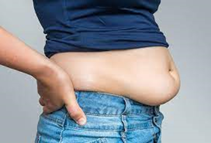
Overweight teenage boy holding his excess abdominal fat

In those with a body mass index (BMI) under 35, intra-abdominal body fat is related to negative health outcomes independent of total body fat.[76] Intra-abdominal or visceral fat has a particularly strong correlation with cardiovascular disease.


BMI and waist measurements are well recognized ways to characterize obesity. However, waist measurements are not as accurate as BMI measurements. Waist measurement (e.g., for BFP standard) is more prone to errors than measuring height and weight (e.g., for BMI standard). BMI will illustrate the best estimate of one's total body fatness, while waist measurement gives an estimate of visceral fat and risk of obesity-related disease.  It is recommended to use both methods of measurements.


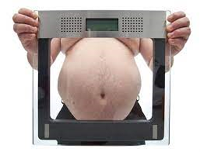
An adult man with abdominal obesity

While central obesity can be obvious just by looking at the naked body (see the picture), the severity of central obesity is determined by taking waist and hip measurements. The absolute waist circumference 102 centimetres (40 in) in men and 88 centimetres (35 in) in women and the waist–hip ratio (>0.9 for men and >0.85 for women are both used as measures of central obesity. A differential diagnosis includes distinguishing central obesity from ascites and intestinal bloating. In the cohort of 15,000 people participating in the National Health and Nutrition Examination Survey (NHANES III), waist circumference explained obesity-related health risk better than BMI when metabolic syndrome was taken as an outcome measure and this difference was statistically significant. In other words, excessive waist circumference appears to be more of a risk factor for metabolic syndrome than BMI. Another measure of central obesity which has shown superiority to BMI in predicting cardiovascular disease risk is the Index of Central Obesity (waist-to-height ratio, WHtR), where a ratio of >=0.5 (i.e. a waist circumference at least half of the individual's height) is predictive of increased risk.  Obesity may also be diagnosed through  the analysis of intra-abdominal fat having the most risk to one's personal health. The increased amount of fat in this region relates to the higher levels of plasma lipid and lipoproteins as per studies mentioned by Eric Poehlman (1998) review. An increasing acceptance of the importance of central obesity within the medical profession as an indicator of health risk has led to new developments in obesity diagnosis such as the Body Volume Index, which measures central obesity by measuring a person's body shape and their weight distribution. The effect of abdominal adiposity occurs not just in those who are obese, but also affects people who are non-obese and it also contributes to insulin sensitivity.


Index of central obesity

Index of Central Obesity (ICO) is the ratio of waist circumference and height first proposed by Parikh et al. in 2007[81] as a better substitute to the widely used waist circumference in defining metabolic syndrome.  The National Cholesterol Education Program Adult Treatment Panel III suggested cutoff of 102 cm (40 in) and 88 cm (35 in) for males and females as a marker of central obesity.The same was used in defining metabolic syndrome. Misra et al. suggested that these cutoffs are not applicable among Indians and the cutoffs be lowered to 90 cm (35 in) and 80 cm (31 in) for males and females. Various race specific cutoffs were suggested by different groups. The International Diabetes Federation defined central obesity based on these various race and gender specific cutoffs. The other limitation of waist circumference is that it the measurement procedure has not been standardized and in children there are no, or few, comparison standards or reference data.


Parikh et al. looked at the average heights of various races and suggested that by using ICO various race- and gender-specific cutoffs of waist circumference can be discarded.[82] An ICO cutoff of 0.53 was suggested as a criterion to define central obesity. Parikh et al. further tested a modified definition of metabolic syndrome in which waist circumference was replaced with ICO in the National Health and Nutrition Examination Survey (NHANES) database and found the modified definition to be more specific and sensitive.


This parameter has been used in the study of metabolic syndrome and cardiovascular disease.


Central obesity in individuals with normal BMI is referred to as normal weight obesity.


Sex differences

There are sex-dependent differences in regional fat distribution.


Males are more susceptible to upper-body fat accumulation, most likely in the belly, due to sex hormone differences. When comparing the body fat of men and women it is seen that men have close to twice the visceral fat as that of pre-menopausal women.


In women, estrogen is believed to cause fat to be stored in the buttocks, thighs, and hips. When women reach menopause and the estrogen produced by ovaries declines, fat migrates from their buttocks, hips, and thighs to their belly.


50% of men and 70% of women in the United States between the ages of 50 and 79 years now exceed the waist circumference threshold for central obesity.


Central obesity is positively associated with coronary heart disease risk in women and men. It has been hypothesized that the sex differences in fat distribution may explain the sex difference in coronary heart disease risk. Even with the differences, at any given level of central obesity measured as waist circumference or waist to hip ratio, coronary artery disease rates are identical in men and women.


Management


A permanent routine of exercise, eating healthily, and, during periods of being overweight, consuming the same number or fewer calories than used will prevent and help fight obesity. A single pound of fat yields approximately 3500 calories of energy (32 000 kJ energy per kilogram of fat), and weight loss is achieved by reducing energy intake, or increasing energy expenditure, thus achieving a negative balance. Adjunctive therapies which may be prescribed by a physician are orlistat or sibutramine, although the latter has been associated with increased cardiovascular events and strokes and has been withdrawn from the market in the US, the UK, the EU, Australia,[105] Canada, Hong Kong] and Thailand.[


A 2006 study published in the International Journal of Sport Nutrition and Exercise Metabolism, suggests that combining cardiovascular (aerobic) exercise with resistance training is more effective than cardiovascular training alone in getting rid of abdominal fat. An additional benefit to exercising is that it reduces stress and insulin levels, which reduce the presence of cortisol, a hormone that leads to more belly fat deposits and leptin resistance.


Self-motivation by understanding the risks associated with abdominal obesity is widely regarded as being far more important than worries about cosmetics. In addition, understanding the health issues linked with abdominal obesity can help in the self-motivation process of losing the abdominal fat. As mentioned above, abdominal fat is linked with cardiovascular disease, diabetes, and cancer. Specifically it's the deepest layer of belly fat (the fat that cannot be seen or grabbed) that poses health risks, as these "visceral" fat cells produce hormones that can affect health (e.g. increased insulin resistance and/or breast cancer risk). The risk increases considering the fact that they are located in the proximity or in between organs in the abdominal cavity. For example, fat next to the liver drains into it, causing a fatty liver, which is a risk factor for insulin resistance, setting the stage for type 2 diabetes. However, visceral fat is more responsive to the circulation of catecholamines.


In the presence of type 2 diabetes, the physician might instead prescribe metformin and thiazolidinediones (rosiglitazone or pioglitazone) as antidiabetic drugs rather than sulfonylurea derivatives. Thiazolidinediones may cause slight weight gain but decrease "pathologic" abdominal fat (visceral fat), and therefore may be prescribed for diabetics with central obesity. Thiazolidinedione has been associated with heart failure and increased cardiovascular risk; so it has been withdrawn from the market in Europe by EMA in 2010.


Low-fat diets may not be an effective long-term intervention for obesity: as Bacon and Aphramor wrote, "The majority of individuals regain virtually all of the weight that was lost during treatment."[113] The Women's Health Initiative ("the largest and longest randomized, controlled dietary intervention clinical trial" found that long-term dietary intervention increased the waist circumference of both the intervention group and the control group, though the increase was smaller for the intervention group. The conclusion was that mean weight decreased significantly in the intervention group from baseline to year 1 by 2.2 kg (P<.001) and was 2.2 kg less than the control group change from baseline at year 1. This difference from baseline between control and intervention groups diminished over time, but a significant difference in weight was maintained through year 9, the end of the study.


Society and culture

Myths

There is a common misconception that spot exercise (that is, exercising a specific muscle or location of the body) most effectively burns fat at the desired location, but this is not the case. Spot exercise is beneficial for building specific muscles, but it has little effect, if any, on fat in that area of the body, or on the body's distribution of body fat. The same logic applies to sit-ups and belly fat. Sit-ups, crunches and other abdominal exercises are useful in building the abdominal muscles, but they have little effect, if any, on the adipose tissue located there.


Colloquialisms

A large central adiposity deposit has been assigned many common use names, including "spare tire", "paunch", and "potbelly". Several colloquial terms used to refer to central obesity, and to people who have it, refer to beer drinking. However, there is little scientific evidence that beer drinkers are more prone to central obesity, despite its being known colloquially as "beer belly", "beer gut", or "beer pot". One of the few studies conducted on the subject did not find that beer drinkers are more prone to central obesity than nondrinkers or drinkers of wine or spirits.   Chronic alcoholism can lead to cirrhosis, symptoms of which include gynecomastia (enlarged breasts) and ascites (abdominal fluid). These symptoms can suggest the appearance of central obesity.


Deposits of excess fat at the sides of one's waistline or obliques are commonly referred to as "love handles".


Economics

Researchers in Copenhagen examined the relationship between waist circumferences and costs among 31,840 subjects aged 50–64 years of age with different waist circumferences. Their study showed that an increase in just an additional centimetre above normal waistline caused a 1.25% and 2.08% rise in health care costs in women and men respectively. To put this in perspective, a woman with a waistline of 95 cm (approx 37.4 in) and without underlying health problems or co-morbidities can incur economic costs that are 22%, or US$397, higher per year than a woman with a normal waist circumference.


Jan Ricks Jennings, MHA, LFACHE

Senior Consultant

Senior Management Resources, LLC

Jan.Jennings@EagleTalons.net

JanJenningsBlog.Blogspot.com


412.913.0636 Cell

724.733.0509 Office


September 24. 2022