Ebola

Ebola, also known as Ebola
virus disease (EVD) and Ebola hemorrhagic fever (EHF), is a viral hemorrhagic
fever in humans and other primates, caused by ebolaviruses.[Symptoms typically
start anywhere between two days and three weeks after becoming infected with
the virus. The first symptoms are usually fever, sore throat, muscle pain, and
headaches. These are usually followed by vomiting, diarrhea, rash and decreased
liver and kidney function, at which point, some people begin to bleed both
internally and externally. The disease kills between 25% and 90% of those
infected – about 50% on average. Death is often due to shock from fluid loss,
and typically occurs between six and 16 days after the first symptoms appear.
Initial treatment of symptoms increases the survival rate considerably compared
to late start. The virus spreads through direct contact with body fluids, such
as blood from infected humans or other animals, or from contact with items that
have recently been contaminated with infected body fluids. There have been no documented cases, either
in nature or under laboratory conditions, of the disease spreading through the
air between humans or other primates. After a person recovers from Ebola, their
semen or breast milk may continue to carry the virus for anywhere between
several weeks to several months. Fruit bats are believed to be the normal
carrier in nature; they can spread the virus without being affected by it. The
symptoms of Ebola may resemble those of several other diseases, including
malaria, cholera, typhoid fever, meningitis, and other viral hemorrhagic
fevers. Diagnosis is confirmed by testing blood samples for the presence of
viral RNA, viral antibodies, or the virus itself. Control of outbreaks requires coordinated medical
services and community engagement,[1] including rapid detection, contact
tracing of those exposed, quick access to laboratory services, care for those
infected, and proper disposal of the dead through cremation or burial. Samples of body fluids and tissues from people
with the disease should be managed with extreme caution. Prevention measures include wearing proper
protective clothing and washing hands when around a person with the disease and
limiting the spread of the disease from infected animals to humans – by wearing
protective clothing while handling potentially infected bushmeat, and by
cooking bushmeat thoroughly before eating it.
An Ebola vaccine was approved in the United States in December 2019. While
there is no approved treatment for Ebola as of 2019, two treatments
(atoltivimab/maftivimab/odesivimab and ansuvimab) are associated with improved
outcomes. Supportive efforts also
improve outcomes. These include oral
rehydration therapy (drinking slightly sweetened and salty water) or giving
intravenous fluids and treating symptoms.
In October 2020,
Atoltivimab/maftivimab/odesivimab (Inmazeb) was approved for medical use in the
United States to treat the disease caused by Zaire ebolavirus. The disease was first identified in 1976, in
two simultaneous outbreaks: one in Nzara (a town in South Sudan) and the other
in Yambuku (the Democratic Republic of the Congo), a village near the Ebola
River, from which the disease takes its name.
Ebola outbreaks occur intermittently
in tropical regions of sub-Saharan Africa.
Between 1976 and 2012, according to the World Health Organization, there
were twenty-four outbreaks of Ebola resulting in a total of 2,387 cases, and
1,590 deaths. The largest Ebola outbreak to date was an epidemic in West Africa
from December 2013 to January 2016, with 28,646 cases and 11,323 deaths. On 29
March 2016, it was declared to no longer be an emergency. Other
outbreaks in Africa began in the Democratic Republic of the Congo in May and 2018.
In July 2019, the World Health
Organization declared the Congo Ebola outbreak a world health emergency. Once the length of time between exposure to
the virus and the development of symptoms (incubation period) is between 2 and
21 days, and usually between 4 and 10 days. However, recent estimates based on
mathematical models predict that around 5% of cases may take longer than 21
days to develop. Symptoms usually begin
with a sudden influenza-like stage characterized by fatigue, fever, weakness,
decreased appetite, muscular pain, joint pain, headache, and sore throat. Zaire
The fever is generally higher than 38.3 °C (101 °F). This is often followed by nausea, vomiting,
diarrhea, abdominal pain, and sometimes hiccups. The combination of severe
vomiting and diarrhea often leads to severe dehydration. Next, shortness of breath and chest pain may
occur, along with swelling, headaches, and confusion.[28] In about half of the
cases, the skin may develop a maculopapular rash, a flat red area covered with
small bumps, five to seven days after symptoms begin. In some cases, internal and external bleeding
may occur. This typically begins five to
seven days after the first symptoms. All infected people show some decreased
blood clotting. Bleeding from mucous
membranes or from sites of needle punctures has been reported in 40–50% of
cases. This may cause vomiting blood, coughing up of blood, or blood in stool. Bleeding into the skin may create petechiae,
purpura, ecchymoses or hematomas (especially around needle injection sites).
Bleeding into the whites of the eyes may also occur. Heavy bleeding is uncommon; if it occurs, it
is usually in the gastrointestinal tract.
The incidence of bleeding into
the gastrointestinal tract was reported to be ~58% in the 2001 outbreak in
Gabon but in the 2014–15 outbreak in the US it was ~18%, due to improved
prevention of disseminated intravascular coagulation.
Recovery
or death
Recovery may begin between
seven and 14 days after first symptoms. Death,
if it occurs, follows typically six to sixteen days from first symptoms and is
often due to shock from fluid loss. In
general, bleeding often indicates a worse outcome, and blood loss may result in
death. People are often in a coma near the end of life. Those who survive often have ongoing muscular
and joint pain, liver inflammation, and decreased hearing, and may have
continued tiredness, continued weakness, decreased appetite, and difficulty
returning to pre-illness weight. Problems with vision may develop. It is recommended that survivors of EVD wear
condoms for at least twelve months after initial infection or until the semen
of a male survivor tests negative for Ebola virus on two separate occasions. Survivors develop antibodies against Ebola
that last at least 10 years, but it is unclear whether they are immune to
additional infections.
Cause
EVD in humans is caused by
four of six viruses of the genus Ebolavirus. The four are Bundibugyo virus
(BDBV), Sudan virus (SUDV), Tai Forest virus (TAFV) and one simply called Ebola
virus (EBOV, formerly Zaire Ebola virus).
EBOV, species Zaire ebolavirus,
is the most dangerous of the known EVD-causing viruses and is responsible for
the largest number of outbreaks. The
fifth and sixth viruses, Reston virus (RESTV) and Bombable virus (BOMV), are
not thought to cause disease in humans, but have caused disease in other
primates. All five viruses are closely
related to Marburg viruses.
Virology
Main
articles: Ebolavirus (taxonomic group) and Ebola virus (specific virus)
Electron
micrograph of an Ebola virus virion
Ebolaviruses contain
single-stranded, non-infectious RNA genomes. Ebolavirus genomes contain seven genes
including 3'-UTR-NP-VP35-VP40-GP-VP30-VP24-L-5'-UTR. The genomes of the five different ebolaviruses
(BDBV, EBOV, RESTV, SUDV and TAFV) differ in sequence and the number and
location of gene overlaps. As with all filoviruses, ebolavirus virions are
filamentous particles that may appear in the shape of a shepherd's crook, of a
"U" or of a "6," and they may be coiled, toroid or
branched. In general, Ebola virions are eighty
nanometers (nm) in width and may be as long as 14,000 nm. Their life cycle is thought to begin with a
virion attaching to specific cell-surface receptors such as C-type lectins,
DC-SIGN, or integrins, which is followed by fusion of the viral envelope with
cellular membranes. The virions taken up
by the cell then travel to acidic endosomes and lysosomes where the viral
envelope glycoprotein GP is cleaved. This processing allows the virus to bind
to cellular proteins enabling it to fuse with internal cellular membranes and
release the viral nucleocapsid. The
Ebolavirus structural glycoprotein (known as GP1,2) is responsible for the
virus' ability to bind to and infect targeted cells. The viral RNA polymerase, encoded by the L
gene, partially uncast the nucleocapsid and transcribes the genes into positive-strand
mRNAs, which are then translated into structural and nonstructural proteins.
The most abundant protein produced is the nucleoprotein, whose concentration in
the host cell determines when L switches from gene transcription to genome
replication. Replication of the viral genome results in full-length,
positive-strand antigenomes that are, in turn, transcribed into genome copies
of negative-strand virus progeny. Newly synthesized
structural proteins and genomes self-assemble and accumulate near the inside of
the cell membrane. Virions bud off from the cell, gaining their envelopes from
the cellular membrane from which they bud. The mature progeny particles then
infect other cells to repeat the cycle. The genetics of the Ebola virus are
difficult to study because of EBOV's virulent characteristics.
Transmission
It is believed that between
people, Ebola disease spreads only by direct contact with the blood or other
body fluids of a person who has developed symptoms of the disease. Body fluids that may contain Ebola viruses
include saliva, mucus, vomit, feces, sweat, tears, breast milk, urine, and
semen. The WHO states that only people who are extremely sick are able to
spread Ebola disease in saliva, and the virus has not been reported to be
transmitted through sweat. Most people spread the virus through blood, feces,
and vomit. Entry points for the virus
include the nose, mouth, eyes, open wounds, cuts, and abrasions. Ebola may be spread through large droplets;
however, this is believed to occur only when a person is extremely sick. This contamination can happen if a person is
splashed with droplets.[61] Contact with surfaces or objects contaminated by
the virus, particularly needles and syringes, may also transmit the infection.
The virus can survive on objects for a few hours in a dried state and can
survive for a few days within body fluids outside of a person.
The Ebola virus may be able
to persist for more than three months in the semen after recovery, which could
lead to infections via sexual intercourse.[6][65][66] Virus persistence in
semen for over a year has been recorded in a national screening program. Ebola may also occur in the breast milk of
women after recovery, and it is not known when it is safe to breastfeed again.
The virus was also found in the eye of one patient in 2014, two months after it
was cleared from his blood. Otherwise,
people who have recovered are not infectious.
The potential for widespread
infections in countries with medical systems capable of observing correct
medical isolation procedures is considered low.
Usually when someone has symptoms
of the disease, they are unable to travel without assistance.
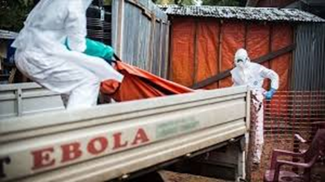
Dead
bodies remain infectious; thus, people managing human remains in practices such
as traditional burial rituals or more modern processes such as embalming are at
risk. Of the cases of Ebola infections in Guinea
during the 2014 outbreak, 69% are believed to have been contracted via
unprotected (or unsuitably protected) contact with infected corpses during
certain Guinean burial rituals.
Healthcare workers treating
people with Ebola are at greatest risk of infection. The risk increases when
they do not have appropriate protective clothing such as masks, gowns, gloves,
and eye protection; do not wear it properly; or handle contaminated clothing
incorrectly. This risk is particularly
common in parts of Africa where the disease mostly occurs, and health systems
function poorly. There has been transmission in hospitals in
some African countries that reuse hypodermic needles. Some health-care centers caring for people
with the disease do not have running water. In the United States the spread to two medical
workers treating infected patients prompted criticism of inadequate training
and procedures.
Human-to-human transmission
of EBOV through the air has not been reported to occur during EVD outbreaks,
and airborne transmission has only been demonstrated in extremely strict
laboratory conditions, and then only from pigs to primates, but not from
primates to primates. Spread of EBOV by
water, or food other than bushmeat, has not been observed. No
spread by mosquitos or other insects has been reported. Other methods of transmission are being
studied.
Airborne transmission among
humans is theoretically possible due to the presence of Ebola virus particles
in saliva, which can be discharged into the air with a cough or sneeze, but
observational data from previous epidemics suggests the actual risk of airborne
transmission is low. Several studies examining airborne
transmission broadly concluded that transmission from pigs to primates could
happen without direct contact because, unlike humans and primates, pigs with
EVD get very high ebolavirus concentrations in their lungs, and not their
bloodstream. Therefore, pigs with EVD
can spread the disease through droplets in the air or on the ground when they
sneeze or cough. By contrast, humans and other primates
accumulate the virus throughout their body and specifically in their blood, but
not very much in their lungs. It is
believed that this is the reason researchers have observed pig to primate
transmission without physical contact, but no evidence has been found of
primates being infected without actual contact, even in experiments where
infected and uninfected primates shared the same air.
Initial
case
Bushmeat
having been smoked in Ghana. In Africa, wild animals including fruit bats are
hunted for food and are referred to as bushmeat.[81][82] In equatorial Africa,
human consumption of bushmeat has been linked to animal-to-human transmission
of diseases, including Ebola.
Although
it is not entirely clear how Ebola initially spreads from animals to humans,
the spread is believed to involve direct contact with an infected wild animal
or fruit bat. Besides bats, other wild
animals that are sometimes infected with EBOV include several species of
monkeys such as baboons, great apes (chimpanzees and gorillas), and duikers (a
species of antelope).
Animals
may become infected when they eat fruit partially eaten by bats carrying the
virus. Fruit production, animal behavior
and other factors may trigger outbreaks among animal populations.
Evidence indicates that both
domestic dogs and pigs can also be infected with EBOV.[86] Dogs do not appear
to develop symptoms when they carry the virus, and pigs are able to transmit
the virus to at least some primates. Although
some dogs in an area in which a human outbreak occurred had antibodies to EBOV,
it is unclear whether they played a role in spreading the disease to people.
Reservoir
The natural reservoir for
Ebola has yet to be confirmed; however, bats are the most likely candidate. Three types of fruit bats (Hypsographic
monstrosus, Epomops franqueti and Myonycteris torquata) were found to possibly
carry the virus without getting sick. As
of 2013, whether other animals participate in its spread is not known. Plants, arthropods, rodents, and birds have
also been considered possible viral reservoirs.
Bats were known to roost in
the cotton factory in which the first cases of the 1976 and 1979 outbreaks were
observed, and they have also been implicated in Marburg virus infections in
1975 and 1980. Of twenty-four plant and
19 vertebrate species experimentally inoculated with EBOV, only bats became
infected. The bats displayed no clinical signs of disease, which is considered
evidence that these bats are a reservoir species of EBOV. In a 2002–2003 survey
of 1,030 animals including 679 bats from Gabon and the Republic of the Congo,
immunoglobulin G (IgG) immune defense molecules indicative of Ebola infection
were found in three bat species; at various periods of study, between 2.2 and
22.6% of bats were found to contain both RNA sequences and IgG molecules
indicating Ebola infection. Antibodies
against Zaire and Reston viruses have been found in fruit bats in Bangladesh,
suggesting that these bats are also potential hosts of the virus and that the
filoviruses are present in Asia Between 1976 and 1998, in 30,000 mammals,
birds, reptiles, amphibians and arthropods sampled from regions of EBOV
outbreaks, no Ebola virus was detected apart from some genetic traces found in
six rodents (belonging to the species Mus sedulous and Praomys) and one shrew (Silvestre
ollula) collected from the Central African Republic. However, further research efforts have not
confirmed rodents as a reservoir. Traces
of EBOV were detected in the carcasses of gorillas and chimpanzees during
outbreaks in 2001 and 2003, which later became the source of human infections.
However, the high rates of death in these species resulting from EBOV infection
make it unlikely that these species represent a natural reservoir for the
virus. Deforestation has been mentioned
as a contributor to recent outbreaks, including the West African Ebola virus
epidemic. Index cases of EVD have often been close to recently deforested
lands.
Pathogenesis
schematic
Like other filoviruses, EBOV replicates very
efficiently in many cells, producing copious amounts of virus in monocytes,
macrophages, dendritic cells, and other cells including liver cells,
fibroblasts, and adrenal gland cells. Viral replication triggers elevated levels of
inflammatory chemical signals and leads to a septic state.
EBOV
is thought to infect humans through contact with mucous membranes or skin
breaks. After infection, endothelial
cells (cells lining the inside of blood vessels), liver cells, and several
types of immune cells such as macrophages, monocytes, and dendritic cells are
the main targets of attack. Following
infection, immune cells carry the virus to nearby lymph nodes where further
reproduction of the virus takes place. From
there the virus can enter the bloodstream and lymphatic system and spread
throughout the body. Macrophages are the
first cells infected with the virus, and this infection results in programmed
cell death. Other types of white blood
cells, such as lymphocytes, also undergo programmed cell death leading to an
abnormally low concentration of lymphocytes in the blood. This contributes to the weakened immune
response seen in those infected with EBOV.
Endothelial cells may be
infected within three days after exposure to the virus. The breakdown of
endothelial cells leading to blood vessel injury can be attributed to EBOV
glycoproteins. This damage occurs due to the synthesis of Ebola virus
glycoprotein (GP), which reduces the availability of specific integrins
responsible for cell adhesion to the intercellular structure and causes liver
damage, leading to improper clotting. The widespread bleeding that occurs in
affected people causes swelling and shock due to loss of blood volume. The
dysfunctional bleeding and clotting commonly seen in EVD has been attributed to
increased activation of the extrinsic pathway of the coagulation cascade due to
excessive tissue factor production by macrophages and monocytes.
After infection, a secreted
glycoprotein, small soluble glycoprotein (saps or GP) is synthesized. EBOV
replication overwhelms protein synthesis of infected cells, and the host immune
defines. The GP forms a trimeric complex, which tethers the virus to the
endothelial cells. The saps forms a dimeric protein that interferes with the signaling
of neutrophils, another type of white blood cell. This enables the virus to
evade the immune system by inhibiting early steps of neutrophil activation.
Immune
system evasion
Filoviral infection also
interferes with proper functioning of the innate immune system. EBOV
proteins blunt the human immune system's response to viral infections by
interfering with the cells' ability to produce and respond to interferon
proteins such as interferon-alpha, interferon-beta, and interferon gamma.
The VP24 and VP35 structural
proteins of EBOV play a key role in this interference. When a cell is infected
with EBOV, receptors located in the cell's cytosol (such as RIG-I and MDA5) or
outside of the cytosol (such as Toll-like receptor 3 (TLR3), TLR7, TLR8 and
TLR9) recognize infectious molecules associated with the virus. On TLR
activation, proteins including interferon regulatory factor three and
interferon regulatory factor 7 trigger a signaling cascade that leads to the
expression of type 1 interferons. The type 1 interferons are then released and
bind to the IFNAR1 and IFNAR2 receptors expressed on the surface of a neighboring
cell. Once interferon has bound to its
receptors on the neighboring cell, the signaling proteins STAT1 and STAT2 are
activated and move to the cell's nucleus.
This triggers the expression of interferon-stimulated genes, which code
for proteins with antiviral properties. EBOV's V24 protein blocks the production of
these antiviral proteins by preventing the STAT1 signaling protein in the neighboring
cell from entering the nucleus. The VP35
protein directly inhibits the production of interferon-beta. By inhibiting these immune responses, EBOV may
quickly spread throughout the body.
Diagnosis
When
EVD is suspected, travel, work history, and exposure to wildlife are key
factors with respect to further diagnostic efforts.
Laboratory
testing
Possible non-specific laboratory indicators
of EVD include a low platelet count; an initially decreased white blood cell
count followed by an increased white blood cell count; elevated levels of the
liver enzymes alanine aminotransferase (ALT) and aspartate aminotransferase
(AST); and abnormalities in blood clotting often consistent with disseminated
intravascular coagulation (DIC) such as a prolonged prothrombin time, partial
thromboplastin time, and bleeding time. Filo virions such as EBOV may be
identified by their unique filamentous shapes in cell cultures examined with
electron microscopy.
The specific diagnosis of
EVD is confirmed by isolating the virus, detecting its RNA or proteins, or
detecting antibodies against the virus in a person's blood. Isolating the virus
by cell culture, detecting the viral RNA by polymerase chain reaction (PCR) and
detecting proteins by enzyme-linked immunosorbent assay (ELISA) are methods
best used in the early stages of the disease and also for detecting the virus
in human remains. Detecting antibodies against the virus is most
reliable in the later stages of the disease and in those who recover IgM
antibodies are detectable two days after symptom onset and IgG antibodies can
be detected six to 18 days after symptom onset. During an outbreak, isolation of the virus
with cell culture methods is often not feasible. In field or mobile hospitals,
the most common and sensitive diagnostic methods are real-time PCR and ELISA. In
2014, with new mobile testing facilities deployed in parts of Liberia, test results
were obtained 3–5 hours after sample submission. In 2015, a rapid antigen test which gives
results in 15 minutes was approved for use by WHO. It is able to confirm Ebola in 92% of those
affected and rule it out in 85% of those not affected.
Differential
diagnosis
Early
symptoms of EVD may be like those of other diseases common in Africa, including
malaria and dengue fever. The symptoms are also similar to those of
other viral hemorrhagic fevers such as Marburg virus disease, Crimean–Congo hemorrhagic
fever, and Lassa fever.
The complete differential
diagnosis is extensive and requires consideration of many other infectious
diseases such as typhoid fever, shigellosis, rickettsia diseases, cholera,
sepsis, borreliosis, EHEC enteritis, leptospirosis, scrub typhus, plague, Q
fever, candidiasis, histoplasmosis, trypanosomiasis, visceral leishmaniasis,
measles, and viral hepatitis, among others.
Non-infectious diseases that
may result in symptoms like those of EVD include acute promyelocytic leukemia, hemolytic
uremic syndrome, snake envenomation, clotting factor deficiencies/platelet
disorders, thrombotic thrombocytopenic purpura, hereditary hemorrhagic
telangiectasia, Kawasaki disease, and warfarin poisoning.
An Ebola vaccine,
rVSV-ZEBOV, was approved in the United States in December 2019. It appears to be fully effective ten days
after being given. It was
studied in Guinea between 2014 and 2016. More than 100,000 people have been vaccinated
against Ebola as of 2019.
Infection
control
VHF
isolation precautions poster
Community
awareness of the benefits on survival chances of admitting cases early is
important for the infected and infection control
Caregivers
British
woman wearing protective gear
People who care for those
infected with Ebola should wear protective clothing including masks, gloves,
gowns and goggles. The U.S. Centers for Disease Control (CDC) recommend that
the protective gear leaves no skin exposed. These measures are also recommended for those
who may handle objects contaminated by an infected person's body fluids. In 2014, the CDC began recommending that
medical personnel receive training on the proper suit-up and removal of
personal protective equipment (PPE); in addition, a designated person,
appropriately trained in biosafety, should be watching each step of these
procedures to ensure they are done correctly. In Sierra Leone, the typical training period
for the use of such safety equipment lasts approximately 12 days. In
2022 in Uganda, lighter personal protection equipment has become available as
well as possibilities to monitor and communicate with patients from windows in
the treatment tents until it is necessary to enter if e.g. a patient's oxygen
levels drop.
Patients and household
members. The infected person should be
in barrier-isolation from other people. All equipment, medical waste, patient waste
and surfaces that may have come into contact with body fluids need to be
disinfected. During the 2014 outbreak,
kits were put together to help families treat Ebola disease in their homes,
which included protective clothing as well as chlorine powder and other
cleaning supplies. Education of
caregivers in these techniques and providing such barrier-separation supplies
has been a priority of Doctors Without Borders.
Disinfection
Ebolaviruses can be
eliminated with heat (heating for 30 to 60 minutes at 60 °C or boiling for five
minutes). To disinfect surfaces, some lipid solvents such as some alcohol-based
products, detergents, sodium hypochlorite (bleach) or calcium hypochlorite
(bleaching powder), and other suitable disinfectants may be used at appropriate
concentrations.
General
population
Education of the public
about the risk factors for Ebola infection and of the protective measures’
individuals may take to prevent infection is recommended by the World Health
Organization. These measures include avoiding direct contact with infected
people and regular hand washing using soap and water.
Bushmeat
Bushmeat, an important
source of protein in the diet of some Africans, should be handled and prepared
with appropriate protective clothing and thoroughly cooked before consumption. Some research suggests that an outbreak of
Ebola disease in the wild animals used for consumption may result in a
corresponding human outbreak. Since 2003, such animal outbreaks have been
monitored to predict and prevent Ebola outbreaks in humans.
Corpses,
burial
If a person with Ebola
disease dies, direct contact with the body should be avoided. Certain burial rituals, which may have
included making various direct contacts with a dead body, require reformulation
so that they consistently maintain a proper protective barrier between the dead
body and the living. Social anthropologists may help find
alternatives to traditional rules for burials.
Transport,
travel, contact
Transportation crews are
instructed to follow a certain isolation procedure, should anyone exhibit
symptoms resembling EVD. As of August 2014, the WHO does not consider
travel bans to be useful in decreasing spread of the disease. In
October 2014, the CDC defined four risk levels used to determine the level of
21-day monitoring for symptoms and restrictions on public activities. In the United States, the CDC recommends that
restrictions on public activity, including travel restrictions, are not
required for the following defined risk levels:
having been in a country with widespread Ebola disease transmission and
having no known exposure (minimal risk); or having been in that country more
than 21 days ago (no risk) encounter with a person showing symptoms; but not
within three feet of the person with Ebola without wearing PPE; and no direct
contact with body fluid having had brief skin contact with a person showing
symptoms of Ebola disease when the person was believed to be not very
contagious (minimal risk) in countries without widespread Ebola disease
transmission: direct contact with a person showing symptoms of the disease
while wearing PPE (minimal risk) contact with a person with Ebola disease
before the person was showing symptoms (no risk). The CDC recommends monitoring for the
symptoms of Ebola disease for those both at "low risk" and at higher
risk.
Laboratory
In laboratories where
diagnostic testing is conducted, biosafety level 4-equivalent containment is
required. Laboratory researchers must be
professionally trained in BSL-4 practices and wear proper PPE.
Isolation
Isolation refers to
separating those who are sick from those who are not. Quarantine refers to
separating those who may have been exposed to a disease until they either show
signs of the disease or are no longer at risk. Quarantine, also known as enforced isolation,
is usually effective in decreasing spread. Governments often quarantine areas where the
disease is occurring or individuals who may transmit the disease outside of an
initial area. In the United States, the law allows
quarantine of those infected with ebolaviruses.
Contact
tracing
Contact
tracing is considered important to contain an outbreak. It involves finding
everyone who had close contact with infected individuals and monitoring them
for signs of illness for 21 days. If any of these contacts comes down with the
disease, they should be isolated, tested and treated. Then the process is
repeated, tracing the contacts' contacts.
Management
While
there is no approved treatment for Ebola as of 2019, two treatments
(atoltivimab/maftivimab/odesivimab and ansuvimab) are associated with improved
outcomes. The U.S. Food and Drug Administration (FDA) advises people to be
careful of advertisements making unverified or fraudulent claims of benefits
supposedly gained from various anti-Ebola products.[139][140]
In
October 2020, the U.S. Food and Drug Administration (FDA) approved
atoltivimab/maftivimab/odesivimab with an indication for the treatment of
infection caused by Zaire ebolavirus.
Standard
support
A
hospital isolation ward in Gulu, Uganda, during the October 2000 outbreak
Treatment
is primarily supportive in nature. Early supportive care with rehydration and
symptomatic treatment improves survival. Rehydration may be via the oral or intravenous
route. These measures may include pain management, and treatment for nausea,
fever, and anxiety] The World Health Organization (WHO) recommends avoiding
aspirin or ibuprofen for pain management, due to the risk of bleeding
associated with these medications.
Blood
products such as packed red blood cells, platelets, or fresh frozen plasma may
also be used. Other regulators of
coagulation have also been tried including heparin in an effort to prevent
disseminated intravascular coagulation and clotting factors to decrease
bleeding. Antimalarial medications and
antibiotics are often used before the diagnosis is confirmed, though there is
no evidence to suggest such treatment helps. Several experimental treatments
are being studied.
Where
hospital care is not possible, the WHO's guidelines for home care have been
relatively successful. Recommendations include using towels soaked in a bleach
solution when moving infected people or bodies and also applying bleach on
stains. It is also recommended that the caregivers wash hands with bleach
solutions and cover their mouth and nose with a cloth.
Intensive
care
Intensive
care is often used in the developed world. This may include maintaining blood
volume and electrolytes (salts) balance as well as treating any bacterial
infections that may develop. Dialysis
may be needed for kidney failure, and extracorporeal membrane oxygenation may
be used for lung dysfunction.
Prognosis
EVD
has a risk of death in those infected of between 25% and 90%. As of September 2014, the average risk of
death among those infected is 50%. The highest risk of death was 90% in the
2002–2003 Republic of the Congo outbreak.
Early admission significantly
increases survival rates
Death,
if it occurs, follows typically six to sixteen days after symptoms appear and
is often due to low blood pressure from fluid loss. Early
supportive care to prevent dehydration may reduce the risk of death.
If
an infected person survives, recovery may be quick and complete. Prolonged
cases are often complicated by the occurrence of long-term problems, such as
inflammation of the testicles, joint pains, fatigue, hearing loss, mood and
sleep disturbances, muscular pain, abdominal pain, menstrual abnormalities,
miscarriages, skin peeling, or hair loss.[25][148] Inflammation and swelling of
the uveal layer of the eye is the most common eye complication in survivors of
Ebola virus disease.[148] Eye symptoms, such as light sensitivity, excess
tearing, and vision loss have been described.
Ebola
can stay in some body parts like the eyes,[150] breasts, and testicles after
infection. Sexual transmission after recovery has been
suspected If sexual transmission occurs
following recovery it is believed to be a rare event.[154] One case of a
condition similar to meningitis has been reported many months after recovery,
as of October 2015.
A
study of forty-four survivors of the Ebola virus in Sierra Leone reported
musculoskeletal pain in 70%, headache in 48%, and eye problems in 14%.
Epidemiology
For
more about specific outbreaks, see List of Ebola outbreaks.
The
disease typically occurs in outbreaks in tropical regions of Sub-Saharan
Africa.[1] From 1976 (when it was first identified) through 2013, the WHO
reported 2,387 confirmed cases with 1,590 overall fatalities.[1][14] The
largest outbreak to date was the Ebola virus epidemic in West Africa, which
caused a large number of deaths in Guinea, Sierra Leone, and Liberia.
1976
Sudan
Cotton
factory in Nzara, South Sudan, where the first outbreak occurred
The
first known outbreak of EVD was identified only after the fact. It occurred
between June and November 1976, in Nzara, South Sudan [44][157] (then part of
Sudan) and was caused by Sudan virus (SUDV). The Sudan outbreak infected 284
people and killed 151. The first identifiable case in Sudan occurred on 27 June
in a storekeeper in a cotton factory in Nzara, who was hospitalized on 30 June
and died on 6 July.[35][158] Although the WHO medical staff involved in the
Sudan outbreak knew that they were dealing with a heretofore unknown disease,
the actual "positive identification" process and the naming of the
virus did not occur until some months later in Zaire.[158]
A CDC worker incinerates
medical waste from Ebola patients in Zaire in 1976.
On 26 August 1976, the
second outbreak of EVD began in Yambuku, a small rural village in Mangala
District in northern Zaire (now known as the Democratic Republic of the
Congo).[159][160] This outbreak was caused by EBOV, formerly designated Zaire
ebolavirus, a different member of the genus Ebolavirus than in the first Sudan
outbreak. The first person infected with the disease was the village school's principal
Mabolo Lokela, who began displaying symptoms on 26 August 1976.[161] Lokela had
returned from a trip to Northern Zaire near the border of the Central African
Republic, after visiting the Ebola River between 12 and 22 August. He was
originally believed to have malaria and was given quinine. However, his
symptoms continued to worsen, and he was admitted to Yambuku Mission Hospital
on 5 September. Lokela died on 8 September 14 days after he began displaying
symptoms.[162][163]
Soon after Lokela's death,
others who had been in contact with him also died, and people in Yambuku began
to panic. The country's Minister of Health and Zaire President Mobutu Sese Seko
declared the entire region, including Yambuku and the country's capital,
Kinshasa, a quarantine zone. No-one was permitted to enter or leave the area,
and roads, waterways, and airfields were placed under martial law. Schools, businesses,
and social organizations were closed.[164] The initial response was led by
Congolese doctors, including Jean-Jacques Muyembe-Tamfum, one of the
discoverers of Ebola. Muyembe took a blood sample from a Belgian nun; this
sample would eventually be used by Peter Piot to identify the previously
unknown Ebola virus.[165] Muyembe was also the first scientist to come into
direct contact with the disease and survive.[166] Researchers from the Centers
for Disease Control and Prevention (CDC), including Piot, co-discoverer of
Ebola, later arrived to assess the effects of the outbreak, observing that
"the whole region was in panic."[167][168][169]
Piot concluded that Belgian
nuns had inadvertently started the epidemic by giving unnecessary vitamin
injections to pregnant women without sterilizing the syringes and needles. The
outbreak lasted 26 days and the quarantine lasted two weeks. Researchers
speculated that the disease disappeared due to the precautions taken by locals,
the quarantine of the area, and discontinuing of the injections.[164]
During this outbreak, Ngoy
Mushola recorded the first clinical description of EVD in Yambuku, where he
wrote the following in his daily log: "The illness is characterized with a
high temperature of about 39 °C (102 °F), hematemesis, diarrhea with blood,
retrosternal abdominal pain, prostration with 'heavy' articulations, and rapid
evolution death after a mean of three days."[170]
The virus responsible for
the initial outbreak, first thought to be the Marburg virus, was later
identified as a new type of virus related to the genus Marburgvirus. Virus
strain samples isolated from both outbreaks were named "Ebola virus"
after the Ebola River, near the first-identified viral outbreak site in
Zaire.[35] Reports conflict about who initially coined the name: either Karl
Johnson of the American CDC team[171] or Belgian researchers.[172]
Subsequently, a number of other cases were reported, almost all centered on the
Yambuku mission hospital or close contacts of another case.[161] In all, 318
cases and 280 deaths (an 88% fatality rate) occurred in Zaire.[173] Although
the two outbreaks were at first believed connected, scientists later realized
that they were caused by two distinct ebolaviruses, SUDV and EBOV.[160]
1995–2014
Cases of Ebola fever in
Africa since 1976
The second major outbreak
occurred in Zaire (now the Democratic Republic of the Congo, DRC), in 1995,
affecting 315 and killing 254.[1]
In 2000, Uganda had an
outbreak infecting 425 and killing 224; in this case, the Sudan virus was found
to be the Ebola species responsible for the outbreak.[1]
In 2003, an outbreak in the
DRC infected 143 and killed 128, a 90% death rate, the highest of a genus
Ebolavirus outbreak to date.[174]
In 2004, a Russian scientist
died from Ebola after sticking herself with an infected needle.[175]
Between April and August
2007, a fever epidemic[176] in a four-village region[177] of the DRC was
confirmed in September to have been cases of Ebola.[178] Many people who
attended the recent funeral of a local village chief died.[177] The 2007
outbreak eventually infected 264 individuals and killed 187.[1]
On 30 November 2007, the
Uganda Ministry of Health confirmed an outbreak of Ebola in the Bundibugyo
District in Western Uganda. After confirming samples evaluated by the United
States National Reference Laboratories and the Centers for Disease Control, the
World Health Organization (WHO) confirmed the presence of a new species of
genus Ebolavirus, which was tentatively named Bundibugyo.[179] The WHO reported
149 cases of this new strain and thirty-seven of those led to deaths.[1]
The WHO confirmed two small
outbreaks in Uganda in 2012, both caused by the Sudan variant. The first
outbreak affected seven people, killing four, and the second affected twenty-four,
killing seventeen.[1]
On 17 August 2012, the
Ministry of Health of the DRC reported an outbreak of the Ebola-Bundibugyo variant
[180] in the eastern region.[181][182] Other than its discovery in 2007, this
was the only time that this variant has been identified as responsible for an
outbreak. The WHO revealed that the virus had sickened fifty-seven people and
killed twenty-nine. The probable cause of the outbreak was tainted bush meat
hunted by local villagers around the towns of Isiro and Viadana.[1][183]
In 2014, an outbreak
occurred in the DRC. Genome-sequencing showed that this outbreak was not related
to the 2014–15 West Africa Ebola virus outbreak, but was the same EBOV species,
the Zaire species.[184] It began in August 2014, and was declared over in
November with 66 cases and 49 deaths.[185] This was the 7th outbreak in the
DRC, three of which occurred during the period when the country was known as
Zaire.[186]
2013–2016 West Africa
Main article: West African
Ebola virus epidemic
Cases and deaths from April
2014 to July 2015 during the 2013–2015 outbreak
In March 2014, the World
Health Organization (WHO) reported a major Ebola outbreak in Guinea, a West
African nation. Researchers traced the
outbreak to a one-year-old child who died in December 2013. The disease rapidly spread to the neighboring
countries of Liberia and Sierra Leone. It was the largest Ebola outbreak ever
documented, and the first recorded in the region. On 8 August 2014, the WHO declared the
epidemic an international public health emergency. Urging the world to offer
aid to the affected regions, its Director-General said, "Countries
affected to date simply do not have the capacity to manage an outbreak of this
size and complexity on their own. I urge the international community to provide
this support on the most urgent basis possible." By mid-August 2014, Doctors Without Borders
reported the situation in Liberia's capital, Monrovia, was
"catastrophic" and "deteriorating daily". They reported
that fears of Ebola among staff members and patients had shut down much of the
city's health system, leaving many people without medical treatment for other
conditions. In a 26 September statement, WHO said,
"The Ebola pathogen infected so many people so quickly, over such a broad
geographical area, for so long."
Intense
contact tracing and strict isolation prevented further spread of the disease in
the countries that had imported cases.
It caused significant
mortality, with a considerable case fatality rate. By the end of the epidemic, 28,616 people had
been infected; of these, 11,310 had died, for a case-fatality rate of 40%. As of 8 May 2016, 28,646 suspected cases and
11,323 deaths were reported; however, the WHO said that these numbers may be
underestimated. Because they work
closely with the body fluids of infected patients, healthcare workers were
especially vulnerable to infection; in August 2014, the WHO reported that 10%
of the dead were healthcare workers.
2014
Ebola virus epidemic in West Africa
In September 2014, it was
estimated that the countries' capacity for treating Ebola patients was
insufficient by the equivalent of 2,122 beds; by December there were a
sufficient number of beds to treat and isolate all reported Ebola cases,
although the uneven distribution of cases was causing serious shortfalls in
some areas. On 28 January 2015, the WHO
reported that for the first time since the week ending 29 June 2014, there had
been fewer than 100 new confirmed cases reported in a week in the three
most-affected countries. The response to the epidemic then moved to a second
phase, as the focus shifted from slowing transmission to ending the epidemic. On 8 April 2015, the WHO reported only thirty
confirmed cases, the lowest weekly total since the third week of May 2014.
On 29 December 2015, 42 days
after the last person evaluated negative for a second time, Guinea was declared
free of Ebola transmission.[203] At that time, a 90-day period of heightened
surveillance was announced by that agency. "This is the first time that
all three countries – Guinea, Liberia and Sierra Leone – have stopped the
original chains of transmission ...", the organization stated in a news
release. A new case was detected in
Sierra Leone on January 2016. However,
the outbreak was declared no longer an emergency on 29 March 2016.
2014
spread outside West Africa
On 19 September, Eric Duncan
flew from his native Liberia to Texas; five days later he began showing
symptoms and visited a hospital but was sent home. His condition worsened and
he returned to the hospital on 28 September, where he died on 8 October. Health
officials confirmed a diagnosis of Ebola on 30 September – the first case in
the United States.
In early October, Teresa
Romero, a 44-year-old Spanish nurse, contracted Ebola after caring for a priest
who had been repatriated from West Africa. This was the first transmission of
the virus to occur outside Africa.[207] Romero assessed negative for the
disease on 20 October, suggesting that she may have recovered from Ebola
infection.[208]
On 12 October, the Centers
for Disease Control and Prevention (CDC) confirmed that a nurse in Texas, Nina
Pham, who had treated Duncan tested positive for the Ebola virus, the first
known case of transmission in the United States. On 15
October, a second Texas health-care worker who had treated Duncan was confirmed
to have the virus. Both of these people
recovered. An unrelated case involved a doctor in New
York City, who returned to the United States from Guinea after working with
Médecins Sans Frontières and tested positive for Ebola on 23 October. The
person recovered and was discharged from Bellevue Hospital on 11 November. On 24 December 2014, a laboratory in Atlanta,
Georgia reported that a technician had been exposed to Ebola.
On 29 December 2014, Pauline
Cafferkey, a British nurse who had just returned to Glasgow from Sierra Leone,
was diagnosed with Ebola at Glasgow's Garnavillo General Hospital. After initial treatment in Glasgow, she was
transferred by air to RAF Northolt, then to the specialist high-level isolation
unit at the Royal Free Hospital in London for longer-term treatment.
2017
Democratic Republic of the Congo
On 11 May 2017, the DRC Ministry of Public
Health notified the WHO about an outbreak of Ebola. Four people died, and four
people survived; five of these eight cases were laboratory-confirmed. A total
of 583 contacts were monitored. On 2 July 2017, the WHO declared the end of the
outbreak.
On 14 May 2018, the World
Health Organization reported that "the Democratic Republic of Congo
reported 39 suspected, probable or confirmed cases of Ebola between 4 April and
13 May, including 19 deaths. Some 393
people identified as contacts of Ebola patients were being followed up. The
outbreak centred on the Bikoro, Iboko, and Wangata areas in Equateur province,
including in the large city of Mbandaka. The DRC Ministry of Public Health
approved the use of an experimental vaccine. On 13 May 2018, WHO
Director-General Tedros Adhanom Ghebreyesus visited Bikoro. Reports emerged that maps of the area were
inaccurate, not so much hampering medical providers as epidemiologists and
officials trying to assess the outbreak and containment efforts. The 2018 outbreak in the DRC was declared over
on 24 July 2018.
2018–2020 Kivu
Main article: Kivu Ebola
epidemic
On 1 August 2018, the
world's 10th Ebola outbreak was declared in North Kivu province of the
Democratic Republic of the Congo. It was the first Ebola outbreak in a military
conflict zone, with thousands of refugees in the area. By November 2018, 200
Congolese had died of Ebola, about half of them from the city of Beni, where
armed groups are fighting over the region's mineral wealth, impeding medical
relief efforts.
By March 2019, this became
the second largest Ebola outbreak ever recorded, with more than 1,000 cases and
insecurity continuing to be the major resistance to providing an adequate
response. As of 4 June 2019, the WHO
reported 2025 confirmed and probable cases with 1357 deaths. In June 2019, two people died of Ebola in neighboring
Uganda.
In July 2019, an infected
man travelled to Goma, home to more than two million people. One week later, on 17 July 2019, the WHO
declared the Ebola outbreak a global health emergency, the fifth time such a
declaration has been made by the organization. A government spokesman said that half of the
Ebola cases are unidentified, and he added that the current outbreak could last
up to three years.
On 25 June 2020, the second
biggest EVD outbreak ever was declared over.
2020 Équateur province
On 1 June 2020, the
Congolese health ministry announced a new DRC outbreak of Ebola in Mbandaka,
Équateur Province, a region along the Congo River. Genome sequencing suggests
that this outbreak, the 11th outbreak since the virus was first discovered in
the country in 1976, is unrelated to the one in North Kivu Province or the
previous outbreak in the same area in 2018. It was reported that six cases had
been identified; four of the people had died. It is expected that more people
will be identified as surveillance activities increase. By 15 June, the case count had increased to 17
with 11 deaths, with more than 2,500 people having been vaccinated. The
11th EVD outbreak was officially declared over on 19 November 2020. By the time the Equator outbreak ended, it had
130 confirmed cases with seventy-five recoveries and 55 deaths.
2021
North
Kivu
On 7 February 2021, the
Congolese health ministry announced a new case of Ebola near Butembo, North
Kivu detected a day before. The case was a 42-year-old woman who had symptoms
of Ebola in Biena on 1 February 2021. A few days after, she died in a hospital
in Butembo. The WHO said that more than seventy people with contact with the
woman had been tracked.
On 11 February 2021, another
woman who had contact with the previous woman died in the same town, and the
number of traced contacts increased to one hundred. A Day after, a third case was detected in
Butembo.
On 3 May 2021, the 12th EVD
outbreak was declared over, resulting in twelve cases and six deaths. Heightened surveillance will continue for 90
days after the declaration, in case of resurgence.
Guinea
In February 2021, Dr Sakoba
Keita, head of Guinea's national health agency confirmed that three people had
died of Ebola in the south-eastern region near the city of Nyerere. A further
five people also evaluated positive. Keita also confirmed more testing was
underway and attempts to trace and isolate further cases had begun. On 14 February, the Guinean government
declared an Ebola epidemic. The outbreak may have started following
reactivation of a latent case in a survivor of an earlier outbreak. As of 4 May 2021, 23 cases were reported, with
no new cases or deaths since 3 April 2021. A 42-day countdown period was
started on 8 May 2021, and on 19 June, the outbreak was declared over.
Côte d’Ivoire
On 14 August 2021, The
Ministry of Health of Cote d’Ivoire confirmed the country's first case of Ebola
since 1994. This came after the Institutes Pasteur in Cote d'Ivoire confirmed
the Ebola Virus Disease in samples collected from a patient, who was
hospitalized in the commercial capital of Abidjan, after arriving from Guinea.
However, on 31 August 2021,
the WHO found that, after further tests in a laboratory in Lyon, the patient
did not have Ebola. The cause of her disease is still being analyzed.
2022
On 23 April 2022, a case of
Ebola was confirmed in the DRC in the Equateur province. The case was a
31-year-old man whose symptoms began on 5 April but did not seek treatment for
over a week. On 21 April, he was admitted to an Ebola treatment center and died
later that day. By 24 May 2022, there were five recorded
deaths in the DRC. On 15 August, the fifth case was buried, and the outbreak
was declared over, 42 days after, on 4 July 2022.
In September 2022, Uganda
reported seven cases infected with the Ebola Sudan strain, but by mid-October
the count had increased to sixty-three. In November 2022 (present), the outbreak in
Uganda continued - still without a vaccine.
Society
and culture
See also: Cultural
Ebolavirus is classified as a biosafety level 4 agent, as well as a Category A
bioterrorism agent by the Centers for Disease Control and Prevention. It has the potential to be weaponized for use
in biological warfare, and was investigated by Biopreparation for such use, but
might be difficult to prepare as a weapon of mass destruction because the virus
becomes ineffective quickly in open air. Fake emails pretending to be Ebola
information from the WHO or the Mexican government have, in 2014, been misused
to spread computer malware. The BBC
reported in 2015 that "North Korean state media has suggested the disease
was created by the U.S. military as a biological weapon.
Literature
Richard Preston's 1995
best-selling book, The Hot Zone, dramatized the Ebola outbreak in Reston,
Virginia.
William Close's 1995 Ebola:
A Documentary Novel of Its First Explosion and 2002 Ebola: Through the Eyes of
the People focused on individuals' reactions to the 1976 Ebola outbreak in
Zaire.
Tom Clancy's 1996 novel,
Executive Orders, involves a Middle Eastern terrorist attack on the United
States using an airborne form of a deadly Ebola virus strain named "Ebola Maringa"
(see Maringa N’S eka).
As the Ebola virus epidemic
in West Africa developed in 2014, a number of popular self-published and
well-reviewed books containing sensational and misleading information about the
disease appeared in electronic and printed formats. The authors of some such
books admitted that they lacked medical credentials and were not technically
qualified to give medical advice. The World Health Organization and the United
Nations stated that such misinformation had contributed to the spread of the
disease.
Other
animals
Wild
animals
Ebola has a high mortality
rate among primates. Frequent outbreaks
of Ebola may have resulted in the deaths of 5,000 gorillas. Outbreaks
of Ebola may have been responsible for an 88% decline in tracking indices of
observed chimpanzee populations in the 420 km2 Losi Sanctuary between 2002 and
2003. Transmission among chimpanzees
through meat consumption constitutes a significant risk factor, whereas contact
between the animals, such as touching dead bodies and grooming, is not.
Recovered gorilla carcasses
have contained multiple Ebola virus strains, suggesting multiple introductions
of the virus. Bodies decompose quickly and carcasses are not infectious after
three to four days. Contact between guerrilla groups is rare, suggesting that
transmission among guerrilla groups is unlikely, and that outbreaks result from
transmission between viral reservoirs and animal populations.
Domestic
animals
In 2012, it was demonstrated
that the virus can travel without contact from pigs to nonhuman primates,
although the same study failed to achieve transmission in that manner between
primates. Dogs may become infected with
EBOV but not develop symptoms. Dogs in some parts of Africa scavenge for food,
and they sometimes eat EBOV-infected animals and the corpses of humans. A 2005
survey of dogs during an EBOV outbreak found that although they remain
asymptomatic, about 32 percent of dogs closest to an outbreak showed a
seroprevalence for EBOV versus nine percent of those farther away. The
authors concluded that there were "potential implications for preventing
and controlling human outbreaks."
In late 1989, Hazelton
Research Products' Reston Quarantine Unit in Reston, Virginia, had an outbreak
of fatal illness amongst certain lab monkeys. This lab outbreak was initially
diagnosed as simian hemorrhagic fever virus (SHFV) and occurred amongst a
shipment of crab-eating macaque monkeys imported from the Philippines.
Hazelton's veterinary pathologist in Reston sent tissue samples from dead
animals to the United States Army Medical Research Institute of Infectious
Diseases (USAMRIID) at Fort Detrick, Maryland, where an ELISA test indicated
the antibodies present in the tissue were a response to Ebola virus and not
SHFV. An electron microscopist from USAMRIID
discovered filoviruses similar in appearance, in crystalloid aggregates and as
single filaments with a shepherd's hook, to Ebola in the tissue samples sent
from Hazelton Research Products' Reston Quarantine Unit. A US Army team headquartered at USAMRIID euthanized
the surviving monkeys and brought all the dead monkeys to Fort Detrick for
study by the Army's veterinary pathologists and virologists, and eventual
disposal under safe conditions. Blood
samples were taken from 178 animal handlers during the incident. Of those, six animal handlers eventually
seroconverted, including one who had cut himself with a bloody scalpel. Despite
its status as a Level‑four
organism and its apparent pathogenicity in monkeys, when the handlers did not
become ill, the CDC concluded that the virus had a very low pathogenicity to
humans.
The Philippines and the
United States had no previous cases of Ebola infection, and upon further
isolation, researchers concluded it was another strain of Ebola, or a new
filovirus of Asian origin, which they named Reston ebolavirus (RESTV) after the
location of the incident. Reston virus (RESTV) can be transmitted to pigs.
Since the initial outbreak it has since
been found in nonhuman primates in Pennsylvania, Texas, and Italy, where the
virus had infected pigs. According to
the WHO, routine cleaning, and disinfection of pig (or monkey) farms with
sodium hypochlorite or detergents should be effective in inactivating the
Reston ebolavirus. Pigs that have been infected with RESTV tend to show
symptoms of the disease.
Research
Researchers looking at
slides of cultures of cells that make monoclonal antibodies. These are grown in
a lab and the researchers are analyzing the products to select the most
promising. As of July 2015, no
medication has been proven safe and effective for treating Ebola. By the time
the Ebola virus epidemic in West Africa began in 2013, there were at least nine
different candidate treatments. Several trials were conducted in late 2014, and
early 2015, but some were abandoned due to lack of efficacy or lack of people
to study.
As
of August 2019, two experimental treatments known as atoltivimab/maftivimab/odesivimab
and ansuvimab were found to be 90% effective.
Diagnostic
tests
The diagnostic tests
currently available require specialized equipment and highly trained personnel.
Since there are few suitable testing centers in West Africa, this leads to
delay in diagnosis. On 29 November
2014, a new 15-minute Ebola test was reported that if successful, "not
only gives patients a better chance of survival, but it prevents transmission
of the virus to other people." The new equipment, about the size of a
laptop and solar powered, allows testing to be done in remote areas. On 29 December 2014, the U.S. Food and Drug
Administration (FDA) approved the Light Mix Ebola Zaire rprt-PCR test for
patients with symptoms of Ebola.
Disease
models
Animal models and non-human
primates are being used to study various aspects of Ebola virus disease.
Developments in organ-on-a-chip technology have led to a chip-based model for
Ebola hemorrhagic syndrome.
Jan Ricks Jennings. MHA,
LFACHE
Senior Consultant
Senior Management Resources,
LLC
Jan.Jennings@EagleTalons.net
JanJenningsBlog.BlogSpot.com
412.913.0636 Cell
724.733.0509 Office
November 20. 2022
Robert F. Kennedy was born
on November 20, 2025. He was
assassinated by Sirhan Sirhan during in Los Angeles during his presidential
campaign.


